Our website has all of the resources and information you might need including Rx forms, shipping labels and informative preparation guides. Don’t see what you need? Give us a call!
Our use of advanced implant planning software allows us to provide you and your patients the absolute best in guided surgery. Our guided surgery staff expert, Brandon Dickerman, is a part-time consultant for Straumann® regarding the coDiagnostiX Guided Surgery System and has lectured and conducted training on it across the country.
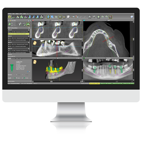
Used by our lab in all of our implant planning, this software allows us to ensure predictable results in every case no matter its complexity.
This user-friendly software helps clinicians in digital treatment planning, diagnostics, and patient communication.
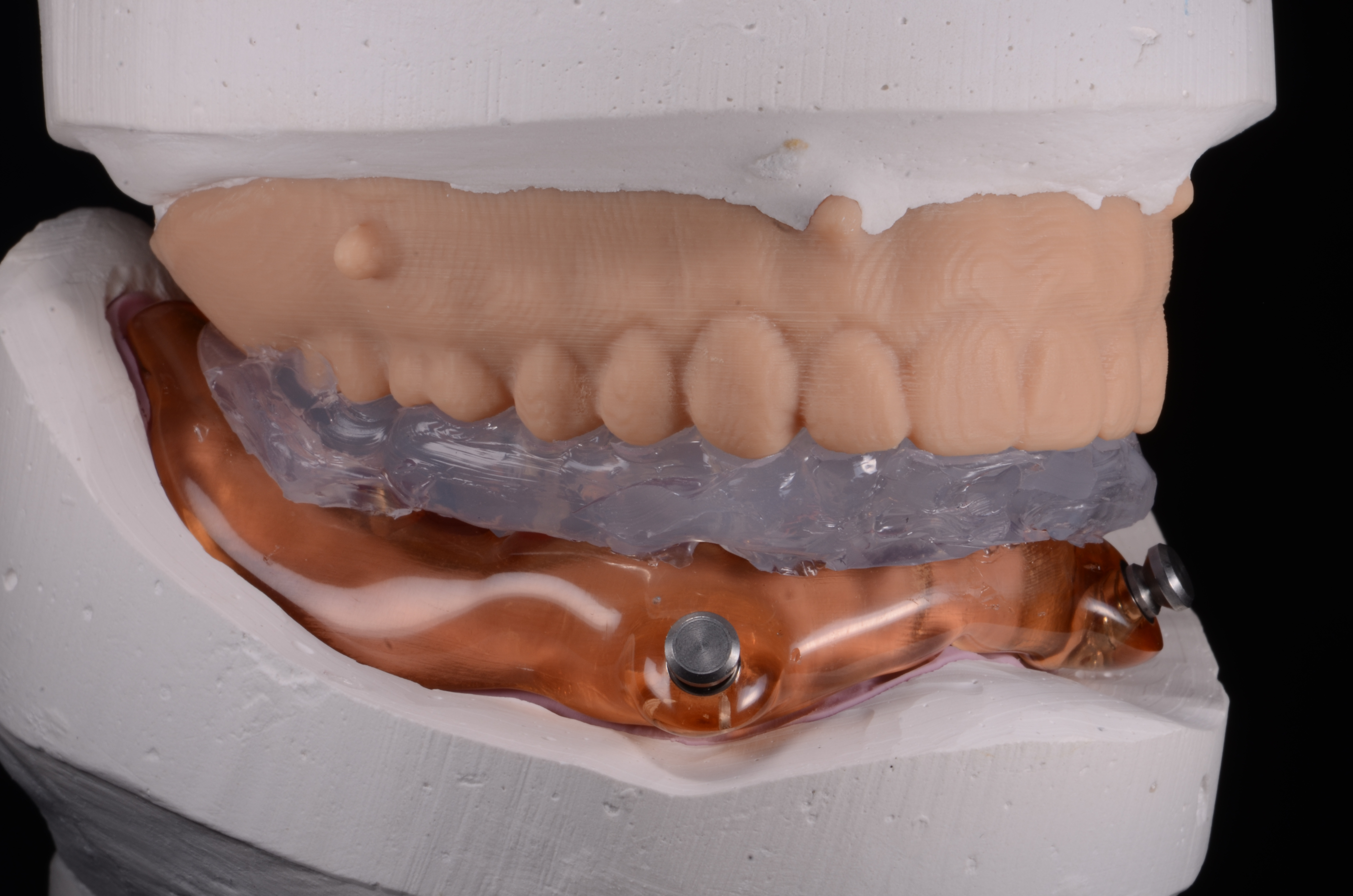
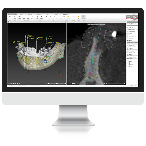
DWOS Synergy™ is a software that allows for a continuous and efficient workflow between our technicians using coDiagnostiX™ and practices using Straumann® CARES® Visual. Synergy allows us to create a digital diagnostic waxup that will be used to plan the placement of implants to ensure truly esthetic results. We will be able to fabricate a laboratory processed pre-surgical provisional restorations with the guided surgical stent. If the intention is to begin to shape the soft tissue from the day of surgery but not load the implant, we can fabricate a custom gingival former that terminates at the soft tissue crest.
All of our technicians at Dickerman Dental Prosthetics are dedicated to simplifying surgeries and reducing patient discomfort and dissatisfaction. When you choose our team of experts you will discover the numerous benefits we provide.
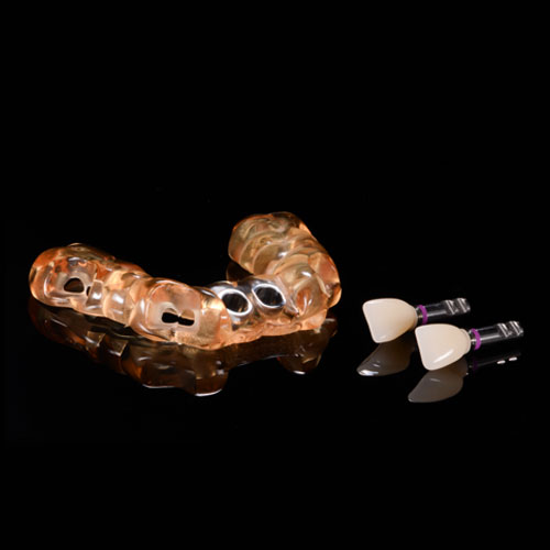
Your patient will appreciate the quality esthetics of our custom provisionals, but its exceptional fit and function will keep them perfectly comfortable throughout the healing process.
Our custom provisionals ensure a perfect fit for your patient, which will decrease the necessary chair time to ensure proper abutment seating.
The provisional will also serve as a blueprint for the final prosthesis. Your patient will be able to provide feedback that will inform the fabrication of the final prosthesis.
When compared to other options, custom healing abutments are more anatomically shaped and biologically advantageous. This provides an easier healing process for the patient.
The CBCT Scanning technique is used to capture all the data needed to fabricate a guided surgical stent. This method utilizes radiographic markers instead of radiographic guides or casts. Please browse the protocols below or view our downloadable PDF.
Order Suremark self stick radiographic markers from us.
Make sure denture fits very well as this will translate into the fit of the surgical guide
If the denture does not fit perfectly, take a wash with light body PVS in the denture with no adhesive
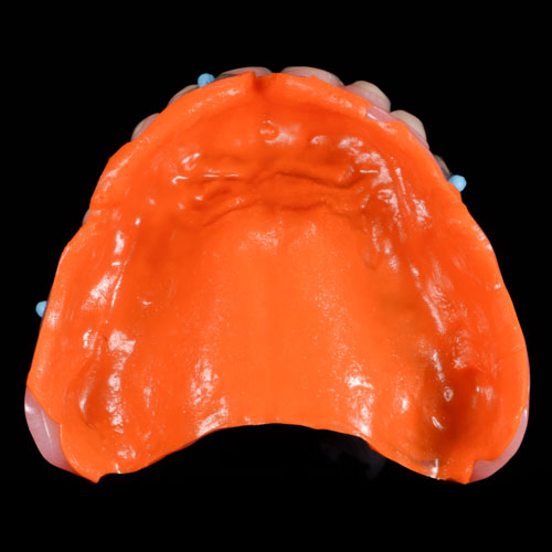
Dry denture and place 3 markers on the buccal flange on different planes and 3 markers on the lingual or palatal for a total of 6 markers

2 CBCT scans will be conducted prior to removing the wash from the denture base or removing the scanning markers
Scan 1: CBCT of the patient with the denture in the mouth biting into occlusion so it is seated properly
Scan 2: CBCT of the denture only with the markers and the wash still in place (no patient).
Please watch this video to see how to set up a CBCT without a patient.
Upload both CBCT scans through our website File Uploader
Common Mistakes
1. Poor fitting denture and either a reline or wash in the denture was not performed at the time of the scan resulting in a poor fitting surgical guide
2. Markers were removed before both scans were performed
3. Markers were not placed on the denture for the scan in the mouth but only for the denture scan
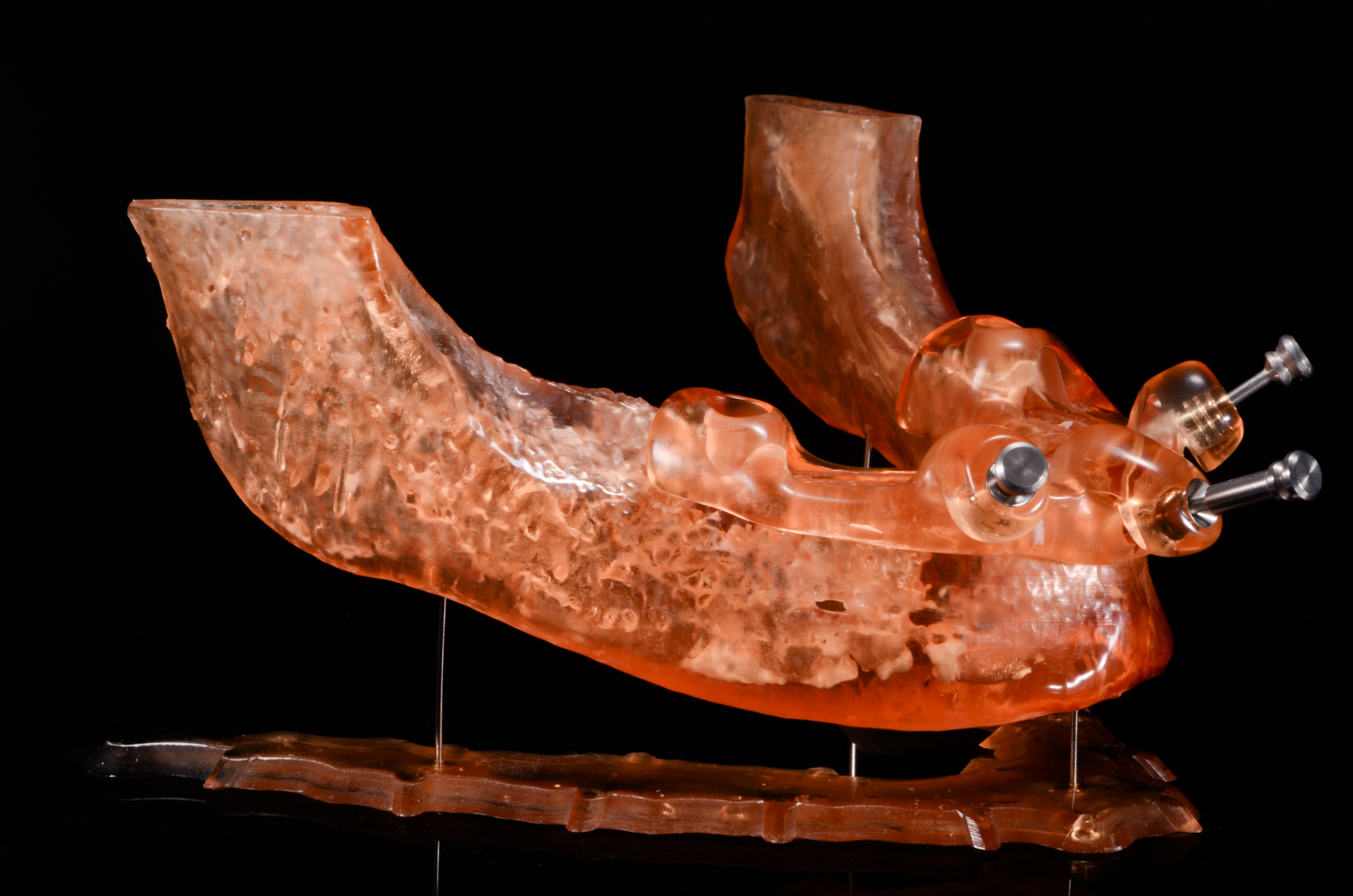
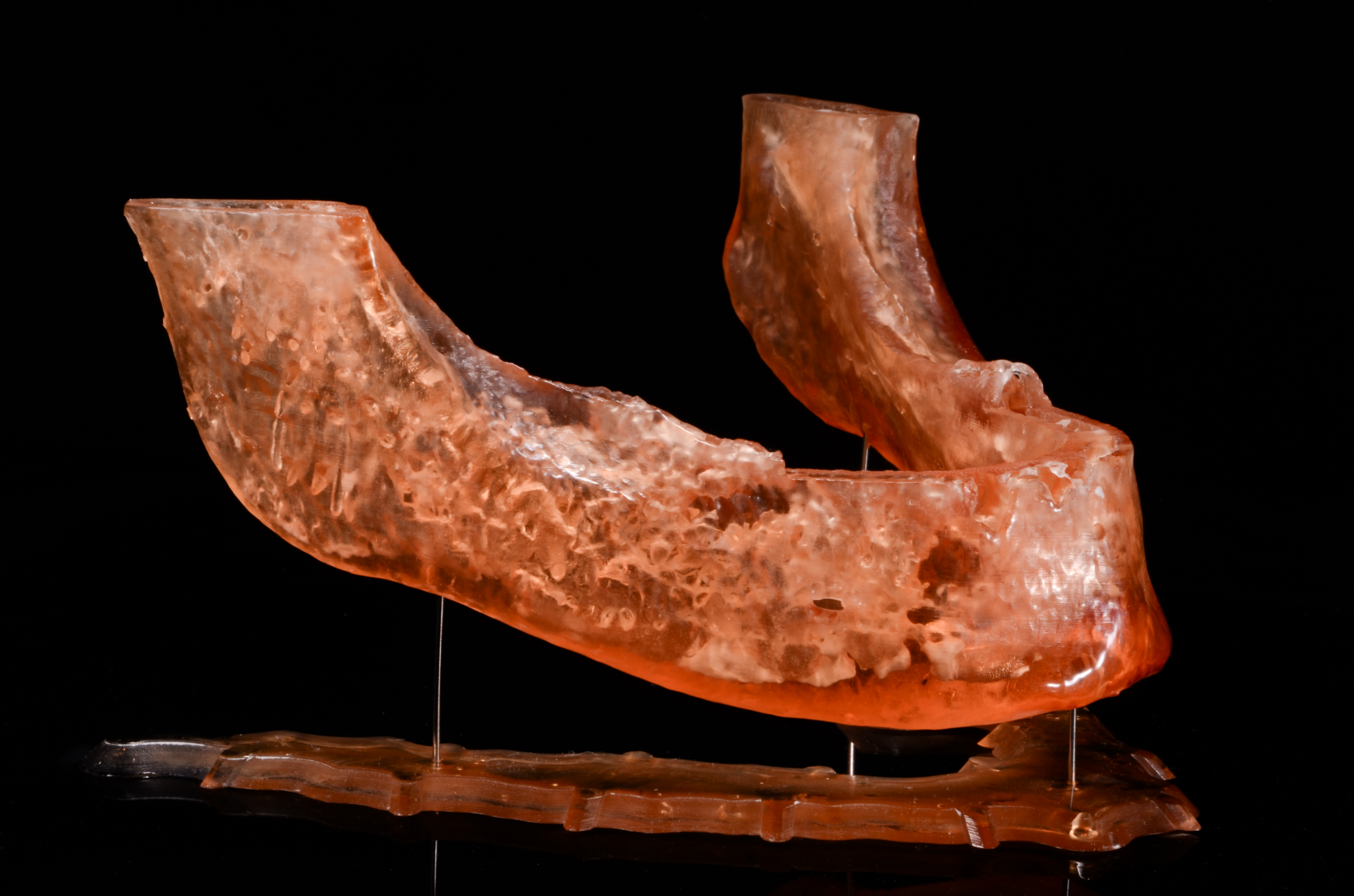
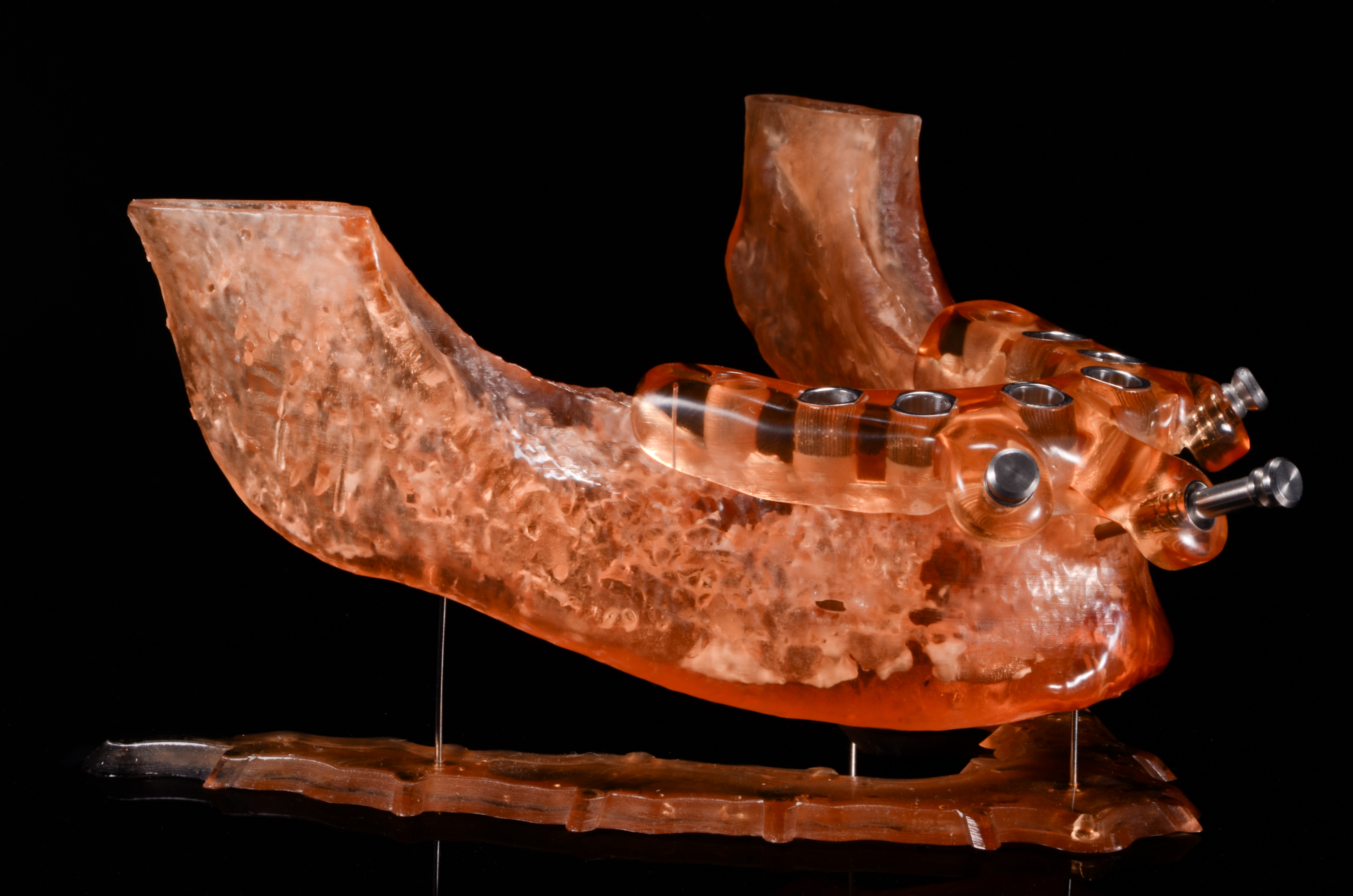
At Dickerman Dental Prosthetics we are well versed in guide fabrication for the edentulous patient including the fabrication of guided bone reduction. We have a workflow in place for fabricating sequential guides starting with a guide for drilling the fixation pin sites prior to reflecting a flap. The second guide is the bone reduction guide that sits directly on the mandibular bone and is supported by the fixation pins. The 3rd and final guide is for drilling the implant osteotomies and is also stabilized on the fixation pins.